June 2026 · 10 min read · Clinical Education
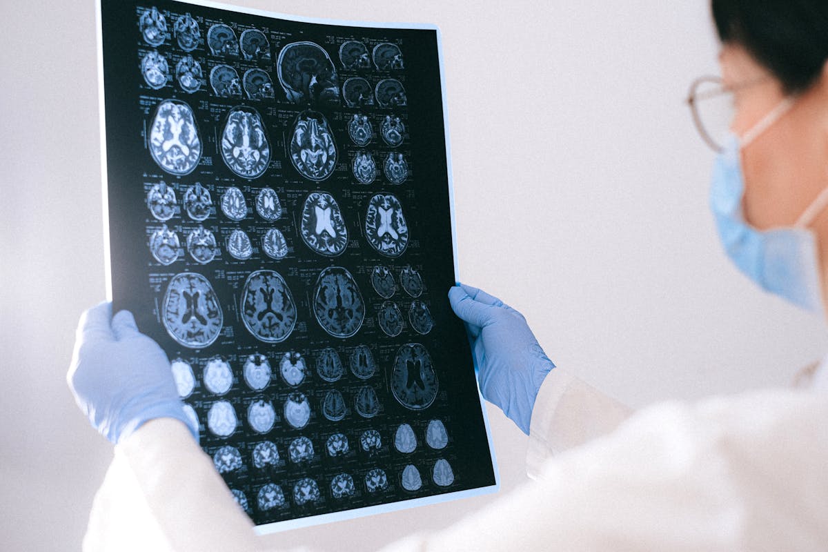
The Anatomy of a Herniated Disc: Why Discs Fail
The intervertebral disc is a remarkable hydrostatic structure. The nucleus pulposus — a gel-like core of proteoglycans and water (80% water in healthy youth) — distributes compressive load across the full cross-sectional area of the disc. Surrounding it, the annulus fibrosus provides tensile containment through 15–25 concentric lamellar rings of collagen fibres, angled alternately at 30° to the vertical. This architecture handles combined compression, bending, and rotation remarkably well — until it doesn't.
Disc failure is not a single event but a process. Degeneration begins with water loss from the nucleus (driven by aging, smoking, and repetitive loading), which reduces hydrostatic pressure. This shifts load from the nucleus to the annulus. Under elevated annular stress, small circumferential tears coalesce into radial fissures. When a radial fissure reaches the outer annulus — where the only disc-nociceptive nerve fibres reside — it becomes painful. When nuclear material follows the fissure outward, a herniation is born.
Primary Biomechanical Causes
1. Cumulative Compressive Loading
Sustained axial compression — from prolonged sitting, heavy manual labour, or occupational vibration (truck drivers, construction workers) — accelerates nucleus dehydration and fatigue microfracture of annular fibres. Seated posture in particular increases L4–L5 intradiscal pressure by 40–90% compared to standing, explaining the high incidence in office and BPO populations.
2. Flexion-Rotation Under Load
The highest-risk movement for disc herniation is trunk flexion combined with rotation while carrying load — the classic "twisted lifting" mechanism. This motion simultaneously: (a) increases posterior intradiscal pressure by up to 300%, (b) partially unlocks the facet joints that normally resist rotation, and (c) concentrates tensile stress on the posterior-lateral annulus — the most common herniation site.
3. Acute Trauma
High-velocity impact (road traffic accidents, falls) can produce acute disc herniation in otherwise healthy discs. In the Philippine context, motorcycle-related trauma and workplace accidents represent a significant acute herniation pathway.
4. Degenerative Cascade
Aging reduces nucleus proteoglycan content, lowering water-binding capacity. By age 50, disc water content has fallen from 80% to 70% in the nucleus; by age 70, to approximately 65%. This alone does not cause herniation, but it creates a mechanically vulnerable disc that herniates at lower loads. The degenerative cascade operates on both a genetic and an environmental axis — twin studies show 60–70% heritability for disc degeneration.
Risk Factor Matrix
| Risk Factor | Mechanism | Relative Risk | Philippine Prevalence |
|---|---|---|---|
| Age 30–50 years | Nucleus dehydration; reduced annular elasticity | Peak incidence | High — dominant working-age population |
| BPO / desk work (>6 hrs/day seated) | Sustained compressive loading; flexed lumbar posture | 2–3× general population | Very high — 1.5M BPO workers |
| Heavy manual labour | Repetitive axial loading + flexion-rotation | 2–4× | High — construction, agriculture, fishing |
| Smoking | Nicotine reduces disc blood supply; impairs proteoglycan synthesis | 1.5–2× | High — 22% adult smoking rate PH |
| Obesity (BMI >30) | Elevated intradiscal pressure; metabolic pro-inflammatory state | 1.5–2× | Rising — 6.4% obesity rate, rapidly increasing |
| Genetic predisposition | Aggrecan, collagen IX, MMP gene variants; 60–70% heritability | Significant | Non-modifiable |
| Prior disc surgery | Adjacent segment disease; altered biomechanics | Elevated at adjacent levels | Growing with surgical volume |
The Molecular Cascade: Why Pain Persists After Herniation
Understanding the molecular sequence from herniation to chronic pain clarifies why PEMF works — and what it is specifically targeting:
- Nucleus pulposus exposure: Herniated nucleus pulposus is immunologically "foreign" to the epidural space — it has no blood supply in healthy disc tissue and is not surveilled by the immune system. When it breaches the annulus, it triggers an autoimmune-like inflammatory response.
- Macrophage infiltration: TNF-α, IL-1β, and IL-6 are released by macrophages engulfing nuclear material. These cytokines directly sensitize adjacent nerve roots, producing radicular pain independent of mechanical compression.
- Nerve root sensitization: Pro-inflammatory mediators lower the depolarization threshold of dorsal root ganglion (DRG) neurons, producing spontaneous ectopic discharges that present as burning, electric, or shooting radicular pain.
- Central sensitization: Persistent afferent input from the sensitized nerve root drives synaptic remodeling in the dorsal horn — long-term potentiation of nociceptive pathways that outlasts the original structural lesion.
How PEMF Interrupts the Herniation-Pain Cascade
PEMF targets the molecular cascade at three intervention points:
Intervention Point 1: Cytokine Suppression
Clinical-grade PEMF reduces TNF-α and IL-1β concentrations in the periradicular space, reducing the chemical irritation of the nerve root. This is the fastest-acting mechanism — patients often report reduced burning quality of radicular pain within the first 3–5 sessions.
Intervention Point 2: Nerve Root Microcirculation
Pulsed fields increase nitric oxide (NO) production and improve capillary perfusion of the compressed nerve root, reducing ischaemic axonal injury that worsens radiculopathy prognosis.
Intervention Point 3: Disc Biology Restoration
A 2026 systematic review (Frontiers in Aging, doi:10.3389/fragi.2026.1840672) confirmed that PEMF activates SIRT1-autophagy pathways in nucleus pulposus cells, promoting clearance of degenerate cellular material and restoration of extracellular matrix components — proteoglycans and type II collagen. This positions PEMF as a disease-modifying intervention, not merely a symptomatic one — a compelling argument for early treatment before disc degeneration becomes irreversible.
The Evidence Base: What the RCTs Show
The evidence for PEMF in disc herniation-related pain is grounded in objective neurophysiological endpoints:
- PMID 23083041 (RCT, n=40, 3-week protocol, lumbar radiculopathy): VAS pain reduction p=0.024; Oswestry Disability Index improvement 9/10 domains (total p<0.001); SSEP latency normalization bilateral p=0.016–0.022; SSEP amplitude improvement bilateral p=0.001–0.002
- PMC11914662 (2025 multicenter RCT, n=91 completers, 5 orthopedic clinics, joint and soft-tissue pain — OA, bursitis, strains, tears): a home-worn 27.12-MHz PEMF wrap used daily for 14 days, against clinician-prescribed standard care (NSAIDs, ice, physical therapy); unblinded. Pain fell 36% vs 10% (p<0.0001); medication use fell 55% vs 12%. It was not a back-pain or disc trial, so its figures cannot be read as results for back pain.
- PMC11775040 (2025 systematic review, 9 RCTs, n=420, chronic LBP): Consistent PEMF superiority over sham and standard care across heterogeneous populations including disc herniation subgroups
- PMC7018371 (Turkish J Physical Medicine & Rehabilitation, cervical disc herniation, n=63): Significant VAS improvement at 12 weeks in PEMF group; safe in routine outpatient use
Conservative vs. Interventional Treatment: Where PEMF Fits
| Treatment | Disc Biology Effect | Pain Mechanism Targeted | Typical Cost (PH) | Risk Level |
|---|---|---|---|---|
| PEMF (clinical grade) | SIRT1-autophagy ECM restoration; cytokine suppression | Cytokine, microcirculation, nerve root sensitization | ₱1,500–₱2,500/session | Very low |
| NSAIDs (oral) | None | Prostaglandin suppression (systemic) | ₱30–₱200/day | GI, renal, CV risk with chronic use |
| Physiotherapy | Indirect — load distribution improvement | Mechanical deloading, motor re-education | ₱800–₱1,500/session | Very low |
| Epidural corticosteroid | None | Targeted anti-inflammatory | ₱8,000–₱25,000/injection | Moderate — infection, bleeding, steroid |
| Discectomy | Mechanical decompression; no regeneration | Structural compression relief | ₱150,000–₱400,000+ | High — surgical, anaesthetic |
PEMF Protocol for Disc Herniation-Related Pain
- Frequency: 10–25 Hz (lumbar); 15–30 Hz (cervical) — lower end for acute, higher for chronic
- Intensity: 30–80 Gauss depending on depth of target tissue and patient tolerance
- Coil placement: centered on affected disc level; secondary applicator over radicular distribution if available
- Session duration: 30–40 minutes
- Session frequency: 2–3 times per week (rest day between sessions)
- Course length: minimum 8 sessions; 12–16 sessions for chronic presentations or Grade III extrusion
- Combination: PEMF → neural mobilization (immediately after) → core stabilization (separate session) for optimal outcome
Prevention: What Clinics Can Offer Beyond Treatment
Disc herniation prevention is a practice-builder for Philippine clinics targeting the BPO and professional workforce:
- Ergonomic assessment: lumbar support positioning, screen height, sit-stand transitions
- Prophylactic PEMF: quarterly 4-session maintenance courses for patients with Grade I disc changes or recurrent axial LBP — reduces progression risk and builds recurring revenue
- Core stability programmes: graduated McGill Big-3 protocol reduces recurrence rate in recovered disc herniation patients
- Smoking cessation support: nicotine cessation alone reduces disc degeneration progression and is a meaningful part of every PEMF treatment plan
Frequently Asked Questions
Can a herniated disc heal on its own?
Spontaneous resorption of extruded disc material does occur — studies show 50–75% of herniations reduce in size over 12 months, with sequestrations having the highest resorption rate. However, this process does not guarantee resolution of symptoms or restoration of disc structure. PEMF accelerates the anti-inflammatory phase of this natural process and may support nucleus pulposus ECM restoration via the SIRT1-autophagy pathway — potentially improving structural outcomes alongside symptomatic relief.
Does sitting always cause disc herniation?
Prolonged, unsupported sitting in lumbar flexion is a significant risk factor, not a guarantee. The key variable is intradiscal pressure over time. Well-supported sitting with lumbar lordosis maintained actually reduces discal pressure compared to flexed sitting. The clinic message is ergonomic posture + periodic deloading + PEMF maintenance, not avoidance of sitting.
Is there a difference between a disc bulge and a disc herniation?
Clinically meaningful distinction: a disc bulge (annular bulging >50% of disc circumference, <3mm beyond the disc border) typically has an intact annulus and often produces axial pain without radiculopathy. A herniation has a focal defect in the annulus and is more likely to produce nerve root compression and radicular symptoms. PEMF is appropriate for both, with protocol parameters adjusted by severity.
PainFree Philippines brings proven clinical PEMF technology — validated in 70+ Israeli clinics serving a population of 9M — to the Philippine market. Request the full clinic operator and investor brief.
Request Investment Brief →